Chondrosarcoma

A 52 year old man is having pain in his right shoulder
Step 3: Imaging and Investigations
Imaging options provide lorem ipsum dolor sit amet, consectetur adipiscing elit. Aliquam nibh est, lobortis eget est quis, hendrerit aliquet urna. Morbi consectetur lacus non metus mollis, porta rut.
X-Ray
 This man has had pain around a joint or bone for more than 6 weeks that is not otherwise explained. In my book, that qualifies a patient for a plain film radiograph. Besides helping us to distinguish between a frozen shoulder and osteoarthritis it will also help to exclude a bone tumour.
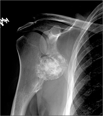
Describe the X-Ray and decide what the diagnosis might be
This is a plain AP radiograph of the shoulder. On the medial side of the humerus there is a surface lesion of bone. Inferiorly the bone is expanded, with medullary and cortical continuity, consistent with a sessile osteochondroma. However, proximal to that is an expanded, irregular bony mass. This mass has a much more aggressive appearance with heterogeneity of the matrix and an irregular border. Otherwise normal appearances. This is highly suggestive (some would go so far as diagnostic) of a chondrosarcoma arising from the surface of an osteochondroma.
MRI
 An MRI scan was also done which confirmed the diagnosis. The scan right shows the thickened cartilage cap on the anterior surface of the lesion. Generally, we accept up to 2cm thickness of the cartilage cap, but this is a little arbitrary. You also need to take into account the age of the patient and whether the cartilage is uniformly thick or has developed one focal area such as in this case.
We elected not to perform a biopsy for this patient. Our group felt that the imaging and clinical features were diagnostic of a chondrosarcoma, and we wanted to avoid the inevitable tissue contamination that occurs from a biopsy. In addition it can be very difficult for pathologists to distinguish between benign cartilage and low grade chondrosarcoma and so the ability of a biopsy to alter the probability that this is a chondrosarcoma is limited, especially when one thinks in Bayesian terms. And so our multidisciplinary conference recommended wide resection. Some centres would have performed a biopsy through an anterior approach, and we don’t think that is wrong, we just feel it is unnecessary. The most important thing is that these decisions are made pre-operatively in a multidisciplinary setting by a group of physicians with extensive experience in bone and soft tissue malignancies