Radiology
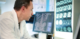
The first step on the way to a diagnosis is often imaging the tumour in some way, whether this be an X-ray, an ultrasound or an MRI. Understanding the role each of these play in the diagnosis of these tumours will help guide your choice of investigation.
Underinvestigation can lead to a missed diagnosis, but unnecessary or unhelpful investigations cost money and subject the patient to unnecessary intervention. This material will help guide you safely between these two dangers.
Systemic Approach to Imaging
The assessment of musculoskeletal tumours relies heavily on the interpretation of radiology studies. All subspecialist units will have a multidisciplinary team that includes a radiologist with subspecialist expertise in musculoskeletal tumours. For bone tumours the plain film X-ray remains the gold standard for diagnosis.
A short guide to reading an X-ray
Background
- Produced by firing X-ray photons at tissue which are absorbed or pass through to a detector plate
- Due to the different densities of air, water, fat and bone an image is produced
- A plain film X-ray will show a bone tumour and the bone’s response to the tumour
Details
- Name
- Date of Birth/Age
- Different diseases occur at different ages
- Clinical Indication
- Side and Views
- Correct side and two views of each bone
- Need to image the whole bone
- Follow the contour of each bone
- Look for signs of tumour, which may be either:
- Lucent areas (black areas in white bone, present due to bone destruction = “lysis”)
- Sclerotic areas (areas that are whiter than expected, due to bone production)
- Look for signs that the lesion is aggressive
- The border is difficult to define
- There is a periosteal reaction on the surface of the bone
- Soft tissue mass
- Fractures may appear in areas of weakened bone
- Look at the soft tissues for any contour abnormality or focal change in density
Choosing an Investigation
When faced with a bone or soft tissue tumour it is important not to over-investigate and waste resources, but also important not to miss serious pathology. For bone lesions a plain film X-ray remains the initial investigation of choice. For soft tissue lesions ultrasound is cheap and easily available so it may be useful as a screening tool, but ultimately the full assessment of a suspicious soft tissue lesion requires an MR scan.
X-Ray
- Initial investigation of choice for any bone lesion
- Image the whole bone in two orthogonal views
- Bone responds very slowly, and the response can be detected on an X-ray
- X-rays give important information about
- Site and Size
- Margins and growth rate
- Periosteal Reactions
- Tumour Matrix
MRI
- MRI is useful for both soft tissue and bone tumours
- Investigation of choice for soft tissue tumours where sarcoma is suspected
- Primary tool for lesions of bone after plain films
CT Scan
- Very good at evaluating osseous lesions, especially the matrix
- Also used to look for metastases to the chest or abdomen
Ultrasound
- Cheap and accessible in the primary care setting
- Gives information on site and size, but is non-specific
- Used to guide biopsy
Bone Scan
- Indicates areas of increased osteoblastic activity
- Guide to the biologic activity of a lesion and metastasis detection
Tips
- Clinical history is essential to narrowing the differential diagnosis.
- Osteosarcoma, also known as Osteogenic Sarcoma, is a tumour that generates an osseous matrix, also known as osteoid. Osteoid is radiodense and appears as white areas on plain films.
- Chondroid tumours produce cartilaginous matrix. On plain films cartilage production looks like punctate ‘dots’ or ‘rings and arcs’.
- Metastases are the most common cause of a bone tumour in patients over 40 years of age, but primary cancer can still occur.
- Pathologic fractures occur very easily through weak bone.
Examples
Well-Differentiated Liposarcoma
- Clinically this mass is highly suspicious as it is large and deep to the deep fascia.
- MRI shows a lesion consistent with a well-differentiated liposarcoma. The lesion has the same signal as fat on all the sequences and has multiple septations.
- These tumours never metastasize, but do have a reputation for local recurrence if not removed in the proper fashion. This patient was managed with surgical resection of the tumour.
Imaging

Dedifferentiated Liposarcoma
- This patient had a mass on his shoulder present for many years. Recently it began to grow rapidly and was causing him significant discomfort.
- MRI shows a lesion that is partly similar to a well-differentiated liposarcoma, but other areas show much higher signal. This is highly suspicious for a high grade sarcoma.
- Core needle biopsy confirmed a dedifferentiated liposarcoma, likely arising in a pre-existing low grade tumour. This is a high grade, dangerous tumour, and so the patient was managed with wide surgical resection of the tumour.
Imaging

Osteosarcoma
- This patient presented with pain and swelling around the upper arm for several months. Examination revealed a hard bony mass.
- Plain X-ray shows a lesion in the humerus producing osteoid. It has poorly defined margins, an aggressive periosteal reaction and is extending into the soft tissues.
- Open biopsy confirmed a high grade osteosarcoma. The patient received chemotherapy and wide surgical resection of the tumour.
Imaging

Ewing Family Tumour
- This patient presented with a painful mass in the shoulder. Examination revealed a large swelling in the scapular region.
- MRI shows a large predominantly soft tissue tumour centered on the scapula. The presence of such a large soft tissue mass strongly suggests a Ewing Family Tumour.
- Core biopsy confirmed a Ewing Family Tumour. The patient was treated with chemotherapy and the scapula lesion was treated with radiation therapy and surgical resection.
Imaging

Synovial Sarcoma
- This patient presented with a growing mass in the upper calf. It was lying deep to the deep fascia and >5 cm in size, so was suspicious for a soft tissue sarcoma.
- MR scans show a soft tissue mass lying deep to the deep fascia. It has signal characteristics suspicious for soft tissue sarcoma.
- Core needle biopsy confirmed synovial sarcoma. The patient was treated with radiation therapy and surgical resection of the tumour.
Imaging

Soft Tissue Sarcoma Lung Metastases
- This patient with known soft tissue sarcoma developed metastases during follow up.
- CT scan of the chest demonstrates innumerable pulmonary nodules.
Imaging

Osteosarcoma Lung Metastasis
- This patient with osteosarcoma developed a lung metastasis, which can be seen on the CT scan of the chest.
- The metastasis is the spiculated mass in the right lung lying anteriorly near the heart.
- A pneumothorax is also present on the right hand side, this can be a complication of lung metastases.
Imaging
